- 3–6 monthsgraft healing when staged
- Same visitas the implant in many cases
- Localanaesthetic for most grafts
- CBCTdecides whether it is needed
What is dental bone grafting?
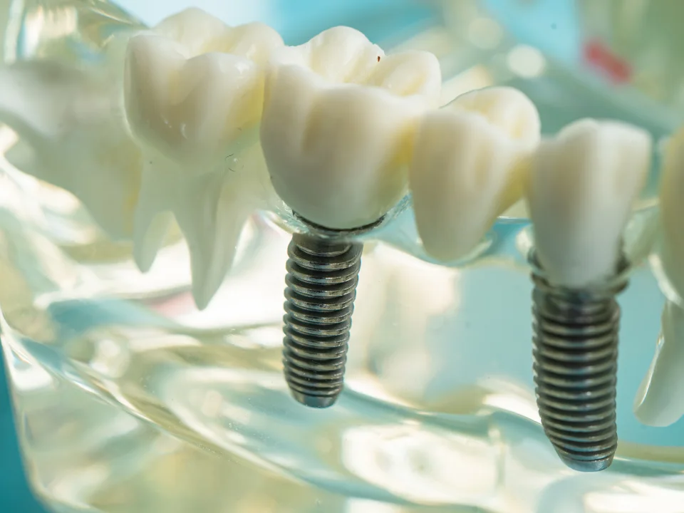
Dental bone grafting in Istanbul is a preparatory procedure in which bone or a bone substitute is added to a deficient area of the jaw, protected while it heals, and allowed to mature into living bone that can hold an implant. It is not a treatment in its own right so much as the foundation for one, and it is planned alongside the implant treatment it serves.
Grafts range from a small amount of material packed into an extraction socket to a substantial augmentation of a ridge that has thinned over years. The material may be your own bone taken from nearby in the jaw, a processed bone substitute, or a combination, often covered by a membrane that keeps soft tissue out while the bone forms.
Why jawbone is lost
Bone exists to support teeth. When a tooth is removed, the bone that held it no longer receives stimulation and begins to resorb, fastest in the first year and continuing slowly thereafter. Gum disease destroys bone around teeth that are still present; long-term denture wear presses on the ridge and accelerates its loss; and infection, cysts or trauma can leave localised defects.
- Resorption after extraction, particularly of the outer wall of the socket
- Periodontitis, which may need gum treatment before any graft
- Years of wearing a full or partial denture
- Infection at the root tip, including after a failed root canal
- Fracture, surgery or a previously failed implant
Types of bone graft used before implants
The technique is chosen to match the shape of the deficiency. Your dentist confirms the approach after examination and CBCT imaging, which shows the width and height of bone at every proposed implant site.
Socket preservation
Graft material is placed into the socket immediately after an extraction to limit the collapse that would otherwise follow. It is quick, done under local anaesthetic, and keeps the option of a straightforward implant a few months later.
Ridge augmentation
For a ridge that has already narrowed, material is added to the outer surface and secured under a membrane, sometimes with small pins or a titanium mesh. Larger defects may use a block of bone fixed with a screw. Healing takes several months before the implant is placed.
Simultaneous grafting
When the deficiency is modest and the implant can still be placed with good stability, grafting is done at the same appointment to cover exposed threads and thicken the outer wall. This avoids a separate healing stage.
Sinus augmentation
In the upper back jaw, height is gained by lifting the sinus membrane and grafting beneath it. This is described separately on the sinus lift page.
The procedure step by step
- Assessment: examination and a CBCT scan to measure the defect and plan the implant position the graft must support.
- Anaesthesia: local anaesthetic for most grafts; sedation for larger augmentations or for anxious patients.
- Access: the gum is gently lifted to expose the deficient area, which is cleaned and prepared.
- Grafting: material is placed and shaped, a membrane is positioned, and the site is closed with sutures.
- Healing: three to six months for a staged graft, during which the material is gradually replaced by your own bone.
- Implant placement: once the CBCT confirms maturation, the implant is placed into the rebuilt ridge.
Healing and aftercare
Swelling is expected for a few days and is more noticeable after larger grafts. The site must be left undisturbed: no pulling the lip to look, no brushing directly on the sutures for the first week, and no pressure from a denture unless it has been adjusted to relieve the area. Prescribed medication and a chlorhexidine rinse protect the site while it seals.
A soft diet on the opposite side for one to two weeks is advised. Smoking is strongly discouraged throughout, since it is one of the main causes of graft failure. Full guidance is in the aftercare guide and is reviewed with you before you fly.
Alternatives to bone grafting
Grafting is not always the answer. Shorter or narrower implants can sometimes be placed in the existing bone without augmentation. Tilted implants, as used in All-on-4, avoid resorbed regions altogether in full-arch cases. For the severely atrophied upper jaw, zygomatic implants anchor in the cheekbone and bypass the need for grafting entirely.
Each alternative has its own indications. The decision weighs the amount of bone missing, the number of teeth to be replaced, the time you can commit and your general health, and it is recorded in your written treatment plan.
Planning grafting as an international patient
A staged graft adds one healing interval but does not necessarily add a trip. A common pattern is: trip one for extraction, grafting or socket preservation; trip two, after three to six months, for implant placement; and trip three for the final crown or bridge. Where the graft is simultaneous with the implant, the schedule stays at two trips.
Each visit needs three to five days so that the site can be checked before travel. Remote follow-up with photographs continues in between. The treatment journey and the costs page explain how staged plans are quoted and scheduled so that there are no surprises.
- Staged graft: typically three trips of 3–5 days each
- Simultaneous graft: two trips, as for a standard implant
- Remote photograph review at set intervals during healing
Risks and what to watch for
The main risks are membrane exposure, infection and partial resorption of the graft, all of which are more likely in smokers and in patients who disturb the site early. Exposure of a small area of membrane is manageable if reported promptly; a fully lost graft can usually be repeated after healing.
Report swelling that increases after day three, pus, a bad taste, sutures that open, visible graft particles in the mouth beyond the first day or two, or a denture that presses on the site. Early contact almost always means a simpler fix.
Related reading
How it works at Habibler Clinic
- AssessmentSend an X-ray or photos; we reply with a written outline.
- Examination & 3D scanClinical check and imaging confirm the plan and timeline.
- TreatmentSessions are scheduled together, in the order agreed.
- Follow-upWritten aftercare and scheduled check-ups.